The Starchild Project
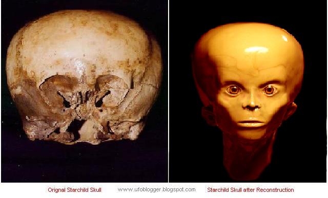
Skulls are humanity’s foremost symbol of death, and a powerful icon in the visual vocabularies of cultures all over the globe. Many strangely “deformed” hominoid skulls have been discovered in Mexico and Peru. One of them, the Starchild skull found in Mexico, is currently the subject of scientific scrutiny and DNA testing.
Thirteen crystal skulls of apparently ancient origin have been found in parts of Mexico, Central America and South America, comprising one of the most fascinating subjects of 20th Century archaeology.
Introduction
In the 1930’s, in a small rural village 100 miles southwest of Chihuahua, Mexico, at the back of a mine tunnel, two mysterious remains were found: a complete human skeleton and a smaller, malformed skeleton.
In late February of 1999, Lloyd Pye was first shown the Starchild skull by its owners. Nameless then, it was a highly anomalous skull. He initially felt it would prove to be a rare genetic deformity of some kind. This skull’s symmetry was astonishing, even more so than the average human. In fact, all of its bones—most of which had human counterparts—were beautifully shaped.
But shaped like what?
Solving many questions that this unusual skull presented became his challenge.
| Front view of the Starchild skull (on the left) and the human skull (on the right). Compare striking differences between depth of eye sockets and shape of temporal area just behind outer edges of eyes. |
Skull Discovery
Sixty to seventy years ago an American girl of Mexican heritage in her late teens (15 to 18) was taken by her parents to visit relatives living in a small rural village 100 miles southwest of Chihuahua, Mexico. The girl was forbidden to enter any of the area’s numerous caves and mine tunnels, but like most teenagers, she went exploring. At the back of a mine tunnel she found a complete human skeleton lying on the ground’s surface.
Beside it, sticking up out of the ground, was a malformed skeletal hand entwined in one of the human skeleton’s upper arms. The girl proceeded to scrape the dirt off a shallow grave to reveal a buried skeleton smaller than the human one and also malformed. She did not specify the type or degree of any of the “malformations.”
The girl recovered both skulls and kept them for the remainder of her life. Upon her death they were passed to an American man, who maintained possession for five years before passing them to the American couple who now control them.
The Mystery Skull
Skull suturing and baby teeth in a detached piece of maxilla (upper jaw and palate) indicate death around 5 years of age. The face is missing from the upper bridge of the nose to the foramen magnum (the hole where the spine enters the skull), but the cranium and most of both eye orbits (the external parts of the sockets) are intact.
This skull’s degree of humanity is at issue because several aspects of its morphology defy categorizing as genetic defect (inherited), congenital deformation (birth defect), or inflicted deformity (cranial binding).
The Human Skull
A human skull assumed to be Amerindian (an Indian from North or South America) because the rear of its cranium exhibits the flattening that results from being carried in infancy on a cradle board. Tooth wear suggests age at death was around 25 years, plus or minus five. Its smallish size and other reduced points of reference indicate it will likely prove to be female.
Binding
Experts suggest the child’s high degree of occipital (rear-skull) deformity would most likely have resulted from the cranial binding practiced by primitive cultures around the world. However, such binding never extends below the inion (the bump at the back of the head) because the human neck begins just below that point. Furthermore, squeezing a skull’s upper bones out of their natural shape leaves them permanently separated, which results in a life-long “soft spot” at the top of the head.
The child’s skull is well-sutured (no soft spot), with none of the distortions normally caused by binding. Furthermore, the extent of rear flattening extends well past the inion, which has become slightly concave.
This indicates a strong force other than binding (i.e. pathology or a natural design) must have caused the occipital’s extensive deformation.
Brain Volume
Though markedly different in shape, the skulls are roughly the same size. However, they exhibit a stunning difference in brain volume. The average volume for a human brain is 1400 cubic centimeters (cc). The volume of the human skull is 1200 cc, typical for a small human. In contrast, the volume of the child’s skull is 1600 cc, which is 200 cc beyond the average for adult humans.
And had it lived to become an adult, its brain capacity would have grown to 1800 cc or more, well beyond the human average.
| The Starchild’s brain volume, contained inside a cranium the size of a smallish human’s, is 1600 cc. A normal human skull has a brain volume around 1400 cubic centimeters. |
In paleoanthropology (the study of ancient animals) a 200 cc increase in brain capacity of a human type creature warrants the naming of an entirely new species. Homo Erectus averages 200 cc more than Homo Habilis; Homo Archaic is 200 cc more than Erectus; Neanderthal is 200 cc more than Archaic.
Thus, this child might well represent an unknown species of human-like beings.
Weight
An average human skull weighs 2.2 pounds (lbs.). The adult’s skull (which is missing its lower jawbone and teeth) weighs 1 lb., 13.4 ounces. Including the child skull’s piece of detached maxilla (upper jaw), it weighs only 13.5 ounces.
Because it is roughly the size of the adult skull, its bone has to be significantly lighter than typical human bone.
Symmetry
The child’s skull has a high degree of symmetry (similarity on both sides). Usually cranial pathologies will cause differences in degree on either side of the head, along with other distortions.
Thus, it is highly unlikely a cranium so clearly aberrant would exhibit such startling symmetry throughout
Sutures
A CAT scan has shown that none of the sutures between the bones in the child’s skull have sealed themselves off from further growth. Nearly all examples of congenital deformity exhibit some degree of premature sealing of cranial sutures. This makes it highly unlikely, if not virtually impossible, for the child’s skull to be the result of deformity. It seems to have grown naturally into the shape is had taken.
The Eyes
Normal human eye sockets have a recessed (5 cm) conical shape with optic nerves and optic fissures at the inner rear quadrant of the cone. The child’s eye sockets have a shallow (3 cm) scalloped shape with optic nerves and optic fissures moved down and away to the inner bottom. Also, the inner surface of both sockets have incredibly subtle terrain shifts that are impossible to explain in any way other than genetic design.
The shape and width of the eye orbits (the outer edges of the sockets) are equally divergent. The adult’s have the vaguely rectangular shape of normal humans, while the child’s are shaped like a lopsided oval. The adult’s are typically rounded along the top of the rectangle, while the upper part of the child’s oval has a clearly definable edge.
The Ears
The child’s ear canals are clearly visible on both sides of its skull. They seem normal in shape and size and angle of entry, but a recent CAT scan revealed that they are larger and have more depth than normal human inner ears. There is no way to know if an external ear was present or what it may have looked like.
The Sinuses
The child had small maxillary (cheek) sinuses but no trace of frontal sinus cavities. While extremely rare, this condition is supposedly known among both humans and primates.
The Foramen Magnum
The foramen magnum is the hole at the base of the skull where the spinal column connects with the brain. In normal humans the foramen is positioned slightly rear of center to balance the hollow-filled front face against the brain-filled occipital area. The extensive reconfiguration of the child’s skull has somehow caused its foramen magnum to be shifted to a central point that provides much better balance between its rear brain area, and its face and forebrain.
The Necks
Typical human neck attachments begin at the inion, the bump in the middle of the occipital bone, and sweep out in a semicircle that reaches to just behind the ears and converges at the foramen magnum. The distance from any part of the semicircle to the foramen opening averages 5 to 6 centimeters.
In the child’s skull a shallow arc extends about 3 centimeters from the foramen hole, while the inion has somehow become slightly concave. Such a drastic reduction in attachment area means the neck supporting the child’s head must have been from 1/2 to 1/3 that of a normal human. Such thin necks are consistently described as hallmarks of certain alien types (Grays), and of Gray-human hybrids.
Chewing Muscles
In the child, the area available for attaching chewing muscles is every bit as reduced as the attachment area for its neck muscles. And though they are called “chewing” muscles, they are actually used for connecting and holding the lower face to the skull.
Based on such a reduced connection area, the amount of mandible (jawbone) these muscles could have secured must have been greatly reduced.
Human-Alien Hybrids
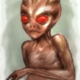
Many abductees and contactees allege that aliens (most often “Grays”) are conducting genetic experiments that produce hybrids between themselves and humans. The results of these unions are consistently described as looking far more human than alien, but with stark bulges in the parietal bones; shallow eye sockets; a greatly reduced lower face; a thin neck able to easily support a well-balanced head; and ears seen as markedly lower and smaller (or missing entirely) relative to human ears.
The eyes of Grays are consistently described and depicted as large black teardrop shapes that wrap horizontally across the middle of the face. If those large orbs are indeed their visual mechanisms, it would argue against the child’s eyes being related to them.
However, in the “Alien Autopsy” film the alien being dissected has the “standard” Gray eyes until the doctor performing the autopsy lifts them off and shows them to actually be dark, flexible coverings like large contact lenses or shades. Underneath those lenses were round, bulging eyes with plenty of white showing around dark irises.
Those eyes would fit quite well in the reduced eye sockets of the child.
The Star Being Legends
These are well-known, well-regarded legends with roots spreading throughout Central and South America. They are pervasive and long-standing (two centuries or more), and in general state that on a regular basis “Star Beings” come down from the heavens and impregnate females in remote, isolated villages. The women carry their “starchildren” to term, then raise them to age six or so.
At that point the Star Beings return to collect their progeny and remove them to places, and for purposes, not clearly outlined in the legends, though improving a stagnated gene pool is often mentioned as a motivation.
The Non-Traditional Scenario
Many “intuitives” and “sensitives” feel the adult skeleton was a female and the child was hers, a human-alien hybrid created by a union between her and a Star Being. Some feel the mother had learned the Star Beings were returning to take her child from her, which she refused to contemplate.
Panic-stricken and filled with dread, she took her child and fled her village, seeking refuge in the hidden mine tunnel. There she killed it and buried it in a shallow grave, leaving one of its hands out of the ground to hold onto.
Then she took a fatal dose of poison and lay down beside her child to die.
DNA Testing
Inside the nucleus of human cells is found nuclear DNA, which is a combination of both parents. Floating outside the nucleus in each of our cells are tiny bits of stray DNA called “mitochondria.” Because mitochondrial DNA (mtDNA) passes solely through females, the first test of the child’s mtDNA will provide a genetic snapshot of its mother. If she was human, that snapshot will say “human.”
However, since the test says nothing about the father, that does not preclude it being a human-alien hybrid. Furthermore, testing might indicate an utterly non-human origin, either by having entirely absent mtDNA or by having a structure markedly different from human mtDNA.
Nothing is likely to be definitive about the origin of the child’s skull until its nuclear DNA can be tested. Because the skull is considered technically “ancient” (over 50 years of age), recovering nuclear DNA will be difficult and costly. Luckily, we have what is most required for such a test, which is teeth. The pulp in teeth resists deterioration better than any other part of the body, so that is where we must look for nuclear DNA.
Worldwide there are only a handful of laboratories capable of sequencing ancient nuclear DNA, and all such processes are time-consuming, highly technical, and very expensive.
Thus, we cannot contract to have this testing done until funding is available to pay for it, but we will announce all such results as soon as they are available.
Mainstream Position:
Pathology–genetic (inherited) or congenital (birth defect)–is the standard explanation for any human-like skull that does not fit the “normal” human mold. In the hands of scientists dedicated to pounding square pegs into the round hole of conventional thinking, pathology can be made to cover virtually any deviation.
In truth, a unique combination of extraordinary pathological disorders is a possible explanation for the many aberrations evident in the child’s skull. Absent overwhelming evidence to the contrary, mainstream science will insist the skull has resulted from nothing more than multiple pathological defects.
This opinion will always dominate any others because of the combined academic credentials of those who will profess it.
This is reality; we all know it.
Points Supporting Non-Earth Origin
The long-standing Star Being legends of Central and South America provide a plausible mechanism for how a highly abnormal skull (relative to humans) might have been biologically created rather than genetically or congenitally malformed, or physically manipulated by deliberate deformation (binding). Such immense deformation across the entire occipital (rear) and parietal (upper side) areas of the skull could not result from binding without deformation being visible in the frontal area, which is not evident.
Birth defects across the entire occipital and parietal areas, while not impossible, seem highly unlikely because of the remarkable symmetry exhibited in all areas of the skull, including those effected by the deformations. The terrain of the bone in the eye sockets contains incredibly subtle indentations and ridges that are perfectly symmetrical in both sockets, which simply have to have been formed by genetic directions rather than by deformations.
The rear deformation extends from the crown to very near the foramen magnum, an area impossible to reach by any binding device due to the thick neck muscles (even in a child) that surround and support the skull-spine connection.
Head binding cannot extend below the inion (the bump at the back of the head). Head binding leaves a gaping opening at the top where skull bones fail to fuse.
The bottom line is that even though the skull’s highly unusual characteristics demand an open-minded approach to it, mainstream science will reject it outright until forced by DNA evidence to do otherwise. Indeed, it could turn out to be nothing more than a butt-ugly kid with an extraordinary combination of cranial deformities never seen before. But it could also have been the result of a human-alien union, or an outright alien with no connection to humanity at all.
Only time and testing will tell which possibility is correct.


 Creators of mankind
 Description of “Tall white aliens”
 Where they came from?
 About hostile civilizations
 The war for the Earth
 “Tall white aliens” about eternal life
 Video: “Nordic aliens”
 Aliens
 Alien encounters
 The aliens base
 UFO
UFO Technology UFO
 Underground civilization
 Ancient alien artifacts
 Military and UFO
 Mysteries and hypotheses
 Scientific facts